Operations on surface meshes
Approximating geometry
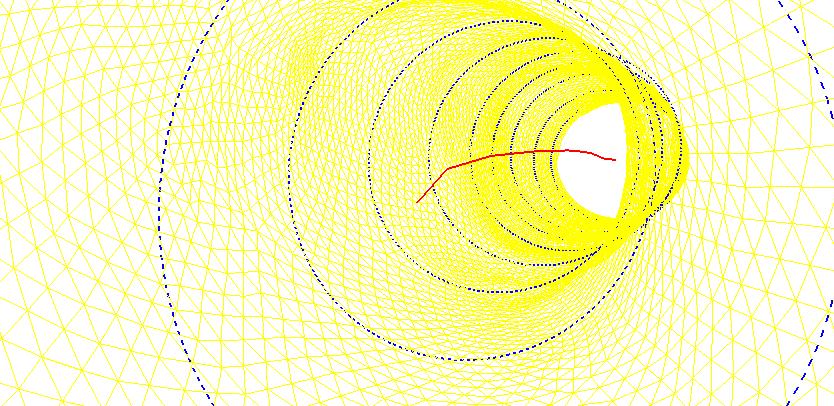
A 3D model of a kinked artery was reconstructed from medical CT scan images and imported into pyFormex as a triangulated surface. The arterial wall surface was then approximated by a sequence of circles with varying diameters and non collinear center points. The figure below shows part of the triangulated surface (yellow), a subset of the approximating circles (blue) and the line connecting the centers of the circles (red). The view is taken from inside the artery.
Stent geometry evaluation
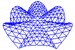
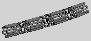
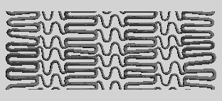
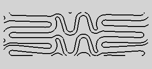
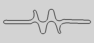
The next figures show four stages in the evaluation of the geometry of a stent device. The first figure is the imported 3D surface, reconstructed from CT scan images. In pyFormex, the tubular stent is unrolled to a planar structure (second figure). This is cut with a plane, producing the circumference, from which the basic cell is taken which is shown in the third figure. Finally, the fourth figure shows the resulting circumference of the opening between the stent material.
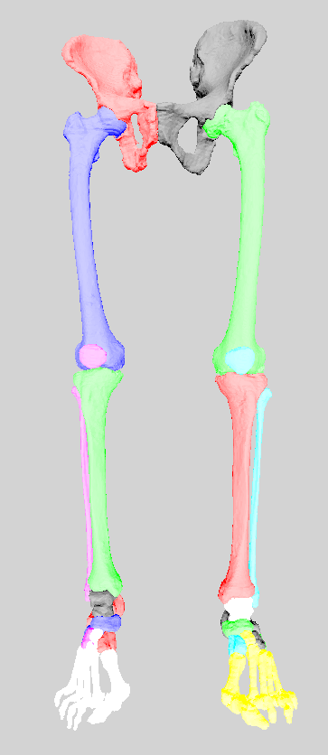
Human skeleton
The individual bones, reconstructed from CT scans, of the lower human skeleton were obtained from the VAKHUM project. The original STL files were converted to GTS format, coarsened, and read into pyFormex, where they were aligned to recreate the skeleton. Colors were added to distinct the bones.